Cricetids (Rodentia, Mammalia) from the Late Miocene Yihachi locality of Gansu, China
QIU Zhu-Ding, Lawrence J. FLYNN, WANG Ban-Yue, LI Lu
Vertebrata Palasiatica
2026, 64 ( 1):
1-25.
DOI: 10.19615/j.cnki.2096-9899.251117
Our purpose in this paper is to describe the hamster-like rodents (Cricetidae) from a Late Miocene age site in Linxia Basin, Gansu Province, and discuss their significance for the changing ecology of central Asia. The micromammal site known as Yihachi was introduced previously (Qiu et al., 2023; Qiu and Li, 2023), when its squirrels were discussed in some detail. We take this opportunity to describe the more abundant cricetids. There are four genera, common Nannocricetus and Sinocricetus plus the less abundant living Mesocricetus. A few specimens represent the high-crowned and lophodont Rhinocerodon. The cricetids and other faunal elements indicate an early Late Miocene age, and the pattern of occurrence of the hamster species is consistent with a picture of a changing paleoenvironment due to increasing effects of the East Asia monsoon system. After the disappearance of older and archaic genera, Yihachi represents growing endemism in the Late Miocene of northern China due to increasing seasonal rain and the declining average temperature.
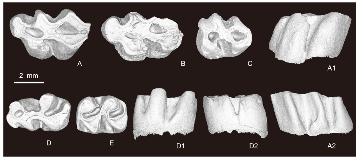
Fig. 8
Molars of Rhinocerodon linxiamys sp. nov. from Yihachi, Gansu
A. r M1 (IVPP V34192, holotype, reversed); B. r M1 (V34193.1, reversed); C. l M3 (V34194.1);
D. l m1 (V34193.2); E. l m2 (V34194.2). A-E. occlusal views; A1, D1. lingual views; A2, D2. buccal views
Extracts from the Article
(Figs. 8, 9)
Holotype IVPP V34192, right M1 (Fig. 8A).
The M1 (Fig. 8A, B) is olive-shaped occlusally. The distinctly larger lingual main cusps have a curved margin lingually, while the buccal ones taper buccally. The anterocone is simple, narrow and crest-like. There are two anterolophules extending from the anterior arm of the protocone and the paracone (protoloph I), respectively. Both anterolophules enclose a long and longitudinal anterofossette with the anterocone, protocone, paracone and the weak protoloph II. The metaloph I is strong and broadly connects with the paracone. The entoloph is thick and strong, joining the paracone. The entoloph and the metaloph I enclose a broad and long posterofossette with the hypocone, metacone, and the weak metaloph II. The posteroloph is short but distinct. The sinus, anterosinus, and mesosinus are broad and deep, and the protosinus is distinct. There are two small enamel lakes in the central and the posterior part of the tooth. The M3 (Fig. 8C) is subrectangular. The anteroloph is pronounced, transverse and confluent with the protocone. There is no lingual anteroloph. The protoloph I is short and thin, joining the anteroloph and dividing the anterosinus into an enclosed anterofossette and an open buccal anterosinus. The entoloph is strong, connecting the hypocone with the paracone, and arranged in an X-form with the protoloph II and the metaloph I. A posterofossette is present, enclosed by the anterior arms of hypocone and metacone and the strong posteroloph. The sinus and mesosinus are broad and deep, the anterosinus is distinct but the posterosinus is indistinct.
The m1 (Fig. 8D) is subrectangular in occlusal surface, with smaller lingual main cusps mesial to the buccal ones. The anteroconid is small and simple. It forms a “sickle-shaped structure” with the protoconid and the metaconid by the connections of the strong anterolophulid and the metalophid I. There is a short buccal branch of anterolophid. The metalophid II is thin and low, joining to the protoconid and closing a round anterofossettid with slight wear. The hypolophid (or anterior arm of entoconid) is thick and meets the ectolophid anteriorly. The ectolophid is prominent and connects the protoconid with the hypoconid. The hypoconid, entoconid, hypolophid and the prominent posterolophid form a “horse-shoe structure”. The sinusid, mesosinusid, and posterosinusid are broad and deep, and the protosinusid is distinct. There is no buccal anterior arm of hypoconid, but an anterobuccal tubercle at the side of the hypoconid. The m2 (Fig. 8E) is rectangular. The anterolophid is marked, extending from the metaconid to the anterobuccal corner of the tooth and joining the base of protoconid. There is no lingual anterolophid. The anterior arm of the protoconid (anterolophulid) is short and joins the anterolophid. A metalophid II is absent, but a spur is present behind the metaconid. The entoconid connects the protoconid to the short hypolophid. The ectolophid joins the junction of the posterior arms of protoconid and hypolophid. The posterolophid is distinct, extending from the hypoconid and tapering to the base of entoconid posterolingually, and closing the posterosinusid. The m2 has two roots.
Other Images/Table from this Article
-
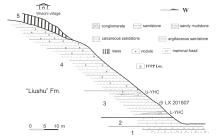 Fig. 1
Section of the fossil locality Yihachi
Fig. 1
Section of the fossil locality Yihachi
-
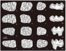 Fig. 2
Molars of Nannocricetus primitivus from Yihachi, Gansu A. l M1 (IVPP V34185.376); B. r M1 (V34185.1); C. l M2 (V34185.377); D. r M2 (V34185.2); E. l M3 (V34185.378); F. r M3 (V34185.3); G. l m1 (V34185.379); H. r m1 (V34185.4); I. l m2 (V34185.380); J. r m2 (V34185.5); K. l m3 (V34185.381); L. r m3 (V34185.6) A-L. occlusal views; A1, G1. lingual views; A2, G2. buccal views
Fig. 2
Molars of Nannocricetus primitivus from Yihachi, Gansu A. l M1 (IVPP V34185.376); B. r M1 (V34185.1); C. l M2 (V34185.377); D. r M2 (V34185.2); E. l M3 (V34185.378); F. r M3 (V34185.3); G. l m1 (V34185.379); H. r m1 (V34185.4); I. l m2 (V34185.380); J. r m2 (V34185.5); K. l m3 (V34185.381); L. r m3 (V34185.6) A-L. occlusal views; A1, G1. lingual views; A2, G2. buccal views
-
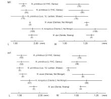 Fig. 3
Size ranges and averages of length and width in the first molars of various species of Nannocricetus from Yihachi, Gansu and other localities in China Measurements of Lantian N. primitivus, N. wuae, Ertemte N. mongolicus and N. qiui are cited from Zhang et al., 2008, 2011; Wu, 1991, and Li et al., 2018, respectively Numbers inside the parentheses are specimen numbers
Fig. 3
Size ranges and averages of length and width in the first molars of various species of Nannocricetus from Yihachi, Gansu and other localities in China Measurements of Lantian N. primitivus, N. wuae, Ertemte N. mongolicus and N. qiui are cited from Zhang et al., 2008, 2011; Wu, 1991, and Li et al., 2018, respectively Numbers inside the parentheses are specimen numbers
-
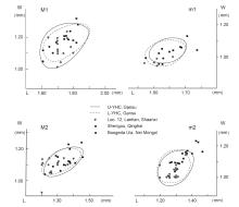 Fig. 4
Scatter diagrams showing the length and width of the first two molars of Nannocricetus primitivus from Yihachi, Gansu and some other localities in northern China Measurements of specimens from Lantian, Shengou and Baogeda Ula are cited from Zhang et al., 2008, Qiu and Li, 2008, 2016
Fig. 4
Scatter diagrams showing the length and width of the first two molars of Nannocricetus primitivus from Yihachi, Gansu and some other localities in northern China Measurements of specimens from Lantian, Shengou and Baogeda Ula are cited from Zhang et al., 2008, Qiu and Li, 2008, 2016
-
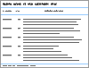 Table 1
Measurements of molars of Nannocricetus primitivus from Yihachi, Gansu (mm)
Table 1
Measurements of molars of Nannocricetus primitivus from Yihachi, Gansu (mm)
-
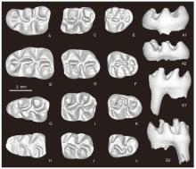 Fig. 5
Molars of Sinocricetus primus sp. nov. from Yihachi, Gansu A. l M1 (IVPP V34186, holotype); B. r M1 (V34188.1); C. l M2 (V34187.1); D. r M2 (V34188.2); E. l M3 (V34187.2); F. r M3 (V34188.3); G. l m1 (V34188.4); H. r m1 (V34187.3); I. l m2 (V34188.5); J. r m2 (V34187.4); K. l m3 (V34188.6); L. r m3 (V34187.5) A-L. occlusal views; A1, G1. lingual views; A2, G2. buccal views
Fig. 5
Molars of Sinocricetus primus sp. nov. from Yihachi, Gansu A. l M1 (IVPP V34186, holotype); B. r M1 (V34188.1); C. l M2 (V34187.1); D. r M2 (V34188.2); E. l M3 (V34187.2); F. r M3 (V34188.3); G. l m1 (V34188.4); H. r m1 (V34187.3); I. l m2 (V34188.5); J. r m2 (V34187.4); K. l m3 (V34188.6); L. r m3 (V34187.5) A-L. occlusal views; A1, G1. lingual views; A2, G2. buccal views
-
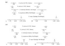 Fig. 6
Size ranges and averages of length and width in the first molars of Sinocricetus primus sp. nov. from Yihachi, Gansu and other species of the genus in China Measurements of S. zdanskyi from Ertemte, S. progressus from Bilike, and S. major from Gaotege are cited from Wu, 1991; Qiu and Storch, 2000; Li, 2010, respectively Numbers inside the parentheses are specimen numbers
Fig. 6
Size ranges and averages of length and width in the first molars of Sinocricetus primus sp. nov. from Yihachi, Gansu and other species of the genus in China Measurements of S. zdanskyi from Ertemte, S. progressus from Bilike, and S. major from Gaotege are cited from Wu, 1991; Qiu and Storch, 2000; Li, 2010, respectively Numbers inside the parentheses are specimen numbers
-
Table 2
Measurements of molars of Sinocricetus primus sp. nov. from Yihachi, Gansu (mm)
-
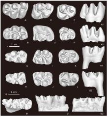 Fig. 7
Molars of Mesocricetus fengi sp. nov. from Yihachi, Gansu A. l M1 (IVPP V34189, holotype); B. r M1 (V34190.2); C. l M2 (V34190.3); D. l M2 (V34191.1, reversed); E. l M3 (V34190.4); F. r M3 (V34190.5); G. l m1 (V34190.6); H. r m1 (V34190.7); I. l m2 (V34190.8); J. r m2 (V34190.9); K. l m3 (V34190.10); L. r m3 (V34190.11), M. right mandibular fragment with m1-2 (V34190.1) A-M. occlusal views; A1, G1, M1. lingual views; A2, G2, M2. buccal views. Scale bars: + for A-L, * for M
Fig. 7
Molars of Mesocricetus fengi sp. nov. from Yihachi, Gansu A. l M1 (IVPP V34189, holotype); B. r M1 (V34190.2); C. l M2 (V34190.3); D. l M2 (V34191.1, reversed); E. l M3 (V34190.4); F. r M3 (V34190.5); G. l m1 (V34190.6); H. r m1 (V34190.7); I. l m2 (V34190.8); J. r m2 (V34190.9); K. l m3 (V34190.10); L. r m3 (V34190.11), M. right mandibular fragment with m1-2 (V34190.1) A-M. occlusal views; A1, G1, M1. lingual views; A2, G2, M2. buccal views. Scale bars: + for A-L, * for M
-
Table 3
Measurements of molars of Mesocricetus fengi sp. nov. from Yihachi, Gansu (mm)
-
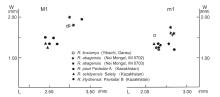 Fig. 9
Scatter diagrams showing length and width in the first molars of Rhinocerodon from Yihachi, Gansu and other species of the genus from Kazakhstan and Nei Mongol Measurements of the specimens of the Kazakhstan species are cited from Zazhigin, 2003
Fig. 9
Scatter diagrams showing length and width in the first molars of Rhinocerodon from Yihachi, Gansu and other species of the genus from Kazakhstan and Nei Mongol Measurements of the specimens of the Kazakhstan species are cited from Zazhigin, 2003
-
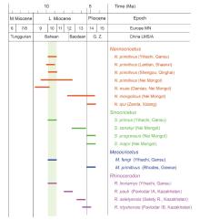 Fig. 10
Biostratigraphic ranges of various species of the cricetid genera present in the Yihachi Fauna G. Z. in the China LMS/A column indicates Gaozhuangian; Shading indicates the proposed ages of Yihachi cricetid composition
Fig. 10
Biostratigraphic ranges of various species of the cricetid genera present in the Yihachi Fauna G. Z. in the China LMS/A column indicates Gaozhuangian; Shading indicates the proposed ages of Yihachi cricetid composition
|
