间型三棱齿象(Trilophodon connexus Hopwood, 1935)属于豕棱齿象类而非嵌齿象
李春晓, 陈津, 王世骐
古脊椎动物学报
2024, 62 ( 1):
33-46.
DOI:10.19615/j.cnki.2096-9899.230917
间型三棱齿象(Trilophodon connexus Hopwood, 1935)长期以来被认为是中国嵌齿象属(Gomphotherium)的一个代表种。然而,由于其下颌联合部与下门齿的形态未知,这一归入存疑。重新研究了来自新疆准噶尔盆地北缘乌伦古河地区哈拉玛盖组的一件此前归为陕西嵌齿象相似种(Gomphotherium cf. G. shensiensis)的下颌。该下颌联合部伸长,呈深槽状,下门齿缺失,因此确定可归入豕棱齿象科(Choerolophodontidae)。进一步将间型三棱齿象的正型标本与其相比较,两者颊齿的关键特征完全一致,包括:高度丘型化,m3伸长,具有四脊,上下颊齿第二脊“人字型”(chevron)很弱,第二脊中附锥与前中心小尖不愈合,釉质褶皱、齿谷中小锥及白垩质发育弱或缺失。因此,间型三棱齿象事实上是一种豕棱齿象类而非嵌齿象。综上所述,暂将其改定为间型“豕棱齿象” (“Choerolophodon” connexus (Hopwood, 1935))。同时,以上特征与北美的索普颌门齿象(Gnathabelodon thorpei)比较接近。此外,在颌门齿象属和间型“豕棱齿象”中,颊齿第二脊呈“人字型”, 釉质褶皱、齿谷中小锥及白垩质发育强这些典型的豕棱齿象属(Choerolophodon)的特征较弱甚至缺失,但m3齿脊数变多,这表明颌门齿象属可能起源于东亚的间型“豕棱齿象”。
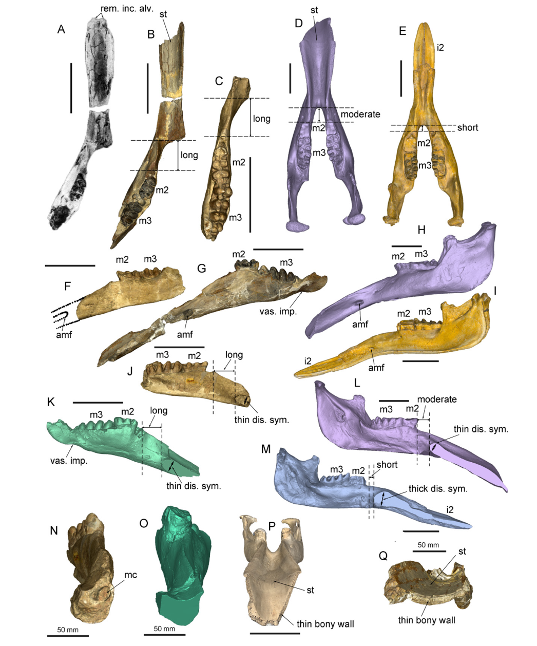
View image in article
Fig. 2
Mandible of “Choerolophodon” connexus and Gnathabelodon, in comparison with Gomphotherium
A, B, G, K, O, Q. “Choerolophodon” connexus, IVPP V8567, from Halamagai Formation, Ulungur region: A. the original photo in Chen (1988:pl. 2, fig. 1); C, F, J, N. “C.” connexus, IVPP RV35015 (cast of the type specimen, PMU-M 3469), from Diaogou, Guanjiashan Formation (formerly Xianshuihe Formation), Xining Basin; D, H, L, P. Gnathabelodon thorpei, FHSU VP18, type specimen, from Ogallah, Kansas, U.S.A., late Clarendonian; note that the distal end of the mandibular symphysis is repaired by plaster; E, I, M. Gomphotherium tassyi, IVPP V22781, from Heijiagou, upper part of Zhangenbao Formation, Zhongning Region
A-E. in dorsal view, showing the deep symphyseal groove and long or moderate distance bewteen the distal end of symphysis and the anterior end of the cheek tooth row; F-I. in left lateral view, showing the tube-like anterior mental foramen; J-M. in right medial view, showing long or moderate distance bewteen the distal end of symphysis and the anterior end of the cheek tooth row, as well as the thin distal end of symphysis; note that K-M were cut along the middle sagital plan from 3D models; N-Q. in distal view, showing the large mandibular channel (N), and thin bony wall of distal symphysis (P, Q); note that O (“C.” connexus, IVPP V8567) was cut from the same position as N (type), which has been broken. Abbreviations: amf. anterior mental foramen; i2. the second lower incisor (mandibular tusk); m2, 3. the second, third lower molar; mc. mandibular channel; rem. inc. alv. remnant of incisor alvoelus; st. symphyseal trough; thick/thin dis. sym. thick/thin distal symphysis; vas. imp. vascular impression for facial artery and vein. Scale bars without notations equal to 20 cm
本文的其它图/表
-
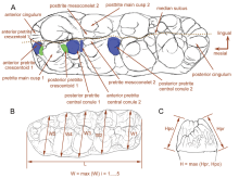 Fig. 1
Terminology and measurements of gomphothere molars A. left m3 of “Choerolophodon” connexus, denoting the terminology of tooth crown; green color, pretrite crescentoids; blue color, pretrite central conules; B. molar crown measurements; C. molar height measurements. Abbreviations: L. length; H. height; Hpo. height of the posttrite side; Hpr. height of the pretrite side; W. width; W1, 2, …, 5. width of the 1st, 2nd, …, 5th loph(id)
Fig. 1
Terminology and measurements of gomphothere molars A. left m3 of “Choerolophodon” connexus, denoting the terminology of tooth crown; green color, pretrite crescentoids; blue color, pretrite central conules; B. molar crown measurements; C. molar height measurements. Abbreviations: L. length; H. height; Hpo. height of the posttrite side; Hpr. height of the pretrite side; W. width; W1, 2, …, 5. width of the 1st, 2nd, …, 5th loph(id)
-
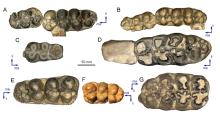 Fig. 3
Cheek teeth of “Choerolophodon” connexus and Gnathabelodon A. “Choerolophodon” connexus, left m2 and m3, IVPP V8567; B. “C.” connexus, left m2 and m3, IVPP RV35015; C. “C.” connexus, right m2, IVPP V31357, from Halamagai Formation, Ulungur region; D. Gnathabelodon thorpei, left m2 and m3, FHSU VP18, type specimen; E. “C.” connexus, right M3, IVPP V8572, from Halamagai Formation, Ulungur region; F. “C.” connexus, left M3, IVPP RV35D49(cast of PMU-M 3045), from Diaogou, Xining Basin; G. Gn. thorpei, right M3, FHSU VP18 Abbreviations: li. lingual side; me. mesial side
Fig. 3
Cheek teeth of “Choerolophodon” connexus and Gnathabelodon A. “Choerolophodon” connexus, left m2 and m3, IVPP V8567; B. “C.” connexus, left m2 and m3, IVPP RV35015; C. “C.” connexus, right m2, IVPP V31357, from Halamagai Formation, Ulungur region; D. Gnathabelodon thorpei, left m2 and m3, FHSU VP18, type specimen; E. “C.” connexus, right M3, IVPP V8572, from Halamagai Formation, Ulungur region; F. “C.” connexus, left M3, IVPP RV35D49(cast of PMU-M 3045), from Diaogou, Xining Basin; G. Gn. thorpei, right M3, FHSU VP18 Abbreviations: li. lingual side; me. mesial side
-
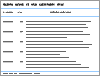 Table 1
Cheek teeth measurements of “Choerolophodon” connexus and Gnathabelodon thorpei (mm)
Table 1
Cheek teeth measurements of “Choerolophodon” connexus and Gnathabelodon thorpei (mm)
-
Table 2
Mandibular measurements of “Choerolophodon” connexus, Gnathabelodon thorpei and Gomphotherium tassyi (mm)
|
