间型三棱齿象(Trilophodon connexus Hopwood, 1935)属于豕棱齿象类而非嵌齿象
Reassessment of Trilophodon connexus Hopwood, 1935 and attributing it to the Choerolophodontidae
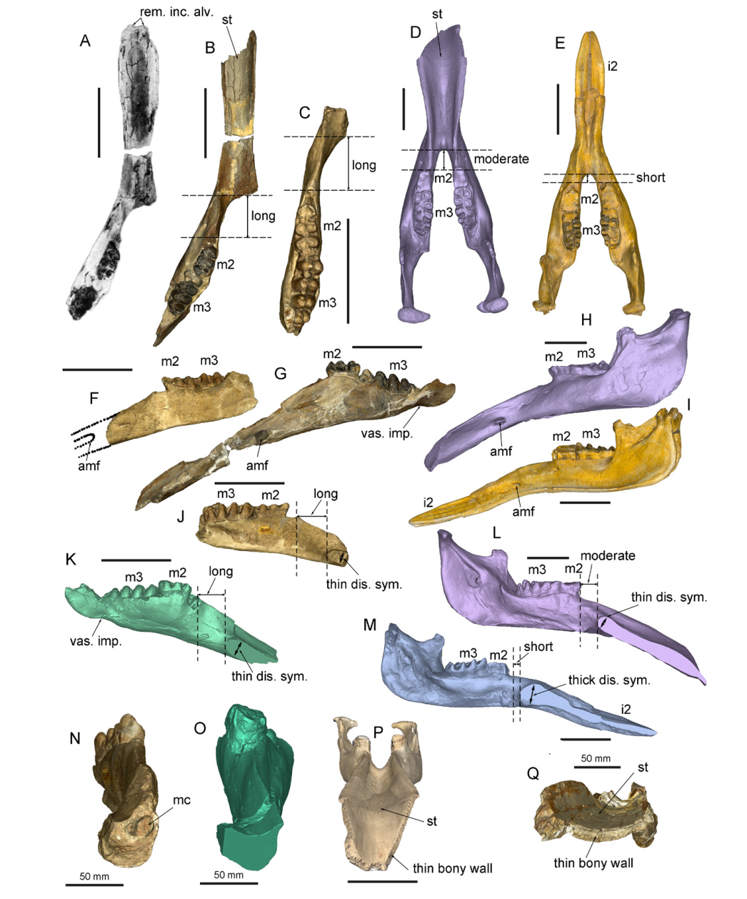
A, B, G, K, O, Q. “Choerolophodon” connexus, IVPP V8567, from Halamagai Formation, Ulungur region: A. the original photo in Chen (
A-E. in dorsal view, showing the deep symphyseal groove and long or moderate distance bewteen the distal end of symphysis and the anterior end of the cheek tooth row; F-I. in left lateral view, showing the tube-like anterior mental foramen; J-M. in right medial view, showing long or moderate distance bewteen the distal end of symphysis and the anterior end of the cheek tooth row, as well as the thin distal end of symphysis; note that K-M were cut along the middle sagital plan from 3D models; N-Q. in distal view, showing the large mandibular channel (N), and thin bony wall of distal symphysis (P, Q); note that O (“C.” connexus, IVPP V8567) was cut from the same position as N (type), which has been broken. Abbreviations: amf. anterior mental foramen; i2. the second lower incisor (mandibular tusk); m2, 3. the second, third lower molar; mc. mandibular channel; rem. inc. alv. remnant of incisor alvoelus; st. symphyseal trough; thick/thin dis. sym. thick/thin distal symphysis; vas. imp. vascular impression for facial artery and vein. Scale bars without notations equal to 20 cm