New fossils of small and medium-sized bovids from the Early Site of Shanshenmiaozui in Nihewan Basin, North China
TONG Hao-Wen, ZHANG Bei, CHEN Xi, WANG Xiao-Min
Vertebrata Palasiatica
2022, 60 ( 2):
134-168.
DOI: 10.19615/j.cnki.2096-9899.220413
Shanshenmiaozui site in Nihewan Basin in North China is a recently discovered Early Pleistocene site which yields rich and diverse mammalian fossils. In the fauna, the small and medium-sized bovid fossils are well represented and can be referred to the following taxa: Spirocerus wongi, Gazella sinensis, Ovis shantungensis and Megalovis piveteaui respectively, among which G. sinensis is the dominate species. S. wongi and G. sinensis are mainly represented by horn-cores and partial skull bones as well as mandibles; in addition, metacarpal and/or metatarsal bones were also recognized for all of the four species. The horn-cores are easy to be identified to the species level, while the dentitions and the postcranial bones underwent a series of examinations and comparisons before getting properly determined and referred to the most approximate taxa. Among the postcranial bones, the metapodials, especially to the metacarpal bones special attentions were paid, which are crucial not only for taxonomic identification, but also for phylogenetic and paleoecological reconstructions; the previously misidentified metapodial specimens in Nihewan fauna were reconsidered in this paper. In the SSMZ fauna, the bovid guild is dominated by Gazella and Bison, which indicates steppe was the most important biome in Nihewan Basin during Early Pleistocene.
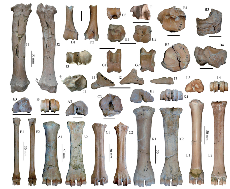
Fig. 4
Postcranial bones of small and medium-sized bovids from SSMZ
A-C. Spirocerus wongi: A1-A3. left Mc III+IV (IVPP V 28655),B1-B4. right naviculo-cuboid+lat-mid+medial cuneiforms (V 28656.2-3),C1-C3. right Mt III+IV (V 28656.4); D-I. Gazella sinensis: D1-D3. partial left humerus (V 28687),E1-E4. left Mc III+IV (V 28688), F. distal epiphysis of left tibia (V 28691),G1-G2. left astragalus (V 28692), H1-H2. left naviculo-cuboid (V 28690), I1-I3. 3rd phalanx (V 28689);J-K. Megalovis piveteaui: J1-J4. left radius (V 28654), K1-K4. right Mc III+IV (V 28695);L1-L4. Ovis shantungensis: left Mc III+IV (V 28694). A1, C1, D1, E1, G1, J1, K1, L1. anterior views;A2, B4, C2, D2, E2, G2, J2, K2, L2. posterior views; A3, B1, C3, E3, H1, J3, K3, L3. proximal views;B2, D3, E4, F, H2, J4, K4, L4. distal views; B3. medial view; I1. lateral view; I2. interdigital view;I3. volar view. The arrows indicate the lateral projection of the radius. The unmarked scale bars equal 20 mm
Extracts from the Article
Mc III+IV: The Mc III+IV (Fig. 4A1-3) is a kind of short but robust one, with the distal end expanded. The cranial aspect is more rounded on the shaft while the caudal surface is more flat. At both the proximal and distal ends the medial side or Mc III looks prominently thicker than the lateral side. In proximal view, there are two large articular facets, the larger facet or magnum trapezoid facet is relatively wide and has a curved edge, which is articulated with the fused 2nd and 3rd carpals; the smaller facet or unciform facet is triangular and articulates with the 4th carpal; between the two facets there exists a groove or depression. The posterior medial tubercle is not developed. In anterior view, the vascular groove (or metacarpal gully) is nearly unobservable and the nutrient foramen is tiny. In posterior view, the shaft has a quite flat surface, except near the proximal end, there is a depression; a small nutrient foramen exists near the distal articular surface. The dimensions are shown in Table 4.
Naviculo-cuboid (IVPP V 28656.1): Except the astragalus and calcaneum, a complete set of right tarsal bone was preserved, which still articulates with the metatarsal bones (V 28656.4) in situ. In proximal view, the articular surface consists of a large trochlear facet with a pair of depressions and a distinct posterior beak-like projection (or internal process) at the medioposterior corner, but the internal process is not developed; a narrow sloped belt-like facet occupies the lateroposterior part, which articulates with the calcaneum; there is a tiny foramen at the bottom of the lateral depression which corresponds with the lateral condyle of the distal trochlea of the astragalus. In distal view, the general outline of the distal surface is that of a rounded quadrilateral with two large anterior articular facets and two small posterior facets; the anteromedial facet is articulated with the fused 2nd and 3rd tarsals; the smallest facet articulates with the 1st tarsal; the other two lateral facets articulated with the Mt III+IV directly. In anterior view, the facet articulated with fused 2nd and 3rd tarsals is higher in position than other distal facets; the calcaneal facet is sloping upward anteroposteriorly. In posterior view, there is a large projection at the posteromedial corner; the posterior surface is not flat and with some small nutrient foramina; there exists a notch between the medial and the calcaneal facets, and the latter extends downward along the back wall (Fig. 4B4). The TD is 43.4 mm, and the APD is 43.8 mm.
The fused 2nd+3rd tarsals (IVPP V 28656.2) (Fig. 4B2-3): Its anatomical position is under the anteromedial portion of the naviculo-cuboid. The proximal articular facet is concave, but the distal facet is convex. Its form is anteroposterior elongated, and its greatest dimension is 29.5 mm.
The 1st tarsal (or medial cuneiform): Short and pillar-like, with facets at both ends; the navicular facet is larger but depressed, and the Mt III facet is smaller but flat. Its anatomical position should be under the beak-like projection of naviculo-cuboid (Fig. 4B2-3).
Mt III+IV (Fig. 4C1-3): In general view, the Mt III+IV has the very proximate length as the Mc III+IV, which is quite different from the common ungulates whose metatarsals are prominently longer than metacarpals belonging to the same individual; furthermore, the distal end expands more sharply relative to the shaft. In proximal view, the proximal surface has a polygon outline, and consists of four articular facets; the two anterior facets are large and kidney-shaped, but the two posterior facets are small and elongated. The mediolateral dimension of the proximal end is slightly larger than the anteroposterior dimension. The two large articular facets meet at the front middle part but diverge posteriorly; both of them have longer anteroposterior dimensions. The anteromedial facet articulates with the fused 2nd and 3rd tarsals; the anterolateral facet articulates with naviculo-cuboid. There are two smaller facets located near the posterior margin. The posterolateral facet has larger mediolateral dimension and articulates with naviculo-cuboid. The small roundish facet articulates with the 1st tarsal. The dimensions are shown in Table 4.
Postcranials: A humerus with partial shaft and complete distal end preserved, the length of the remained part is 73.9 mm (IVPP V 28687) (Fig. 4D1-3). In anterior view, the indentation (radial or coronoid fossa) above the articular surface is shallow; the medial edge of the trochlea is curved and with the upper portion inclined laterally; at the lateral two-fifths part of the trochlear surface, there exists a sagittal ridge whose lower part is nearly vertical but with the upper part slightly inclined laterally; the lowest part of the distal articular surface occurs at the lateral side; there is a longitudinal trench at the middle part of the articular surface; below the articular surface at the medial side, the medial epicondyle can be observed. In posterior view, the shaft has a very straight medial margin and a nutrient foramen at the lateral aspect can be seen; the olecranon fossa at the distal end is very deep; the medial epicondyle extends much more downward than the lateral one. The distal APD is 27.3 mm; the distal TD is 29.7 mm.
Mc III+IV: A complete left Mc III+IV (IVPP V 28688) (Fig. 4E1-4). In general, the bone is very slender; the cranial aspect is more rounded on the shaft and the caudal aspect is much flatter and slightly concave at the upper part; the distal epiphysis is not fused yet. In proximal view, there are two articular facets, the larger one or magnum trapezoid facet is prominently wider and deeper, which articulates with the fused 2nd and 3rd carpals; the smaller one or unciform facet is oval-like and is the articular surface for 4th carpal; between the two facets there exists a narrow ridge; no nutrient foramen can be observed at the proximal end (Fig. 4E3). In anterior view, a prominent tubercle (extensor carpi radialis insertion: Gentry, 1966: fig. 12) occurs at the cranial aspect of the proximal end; no vascular groove (or metacarpal gully) can be observed, while the nutrient foramen is clear but small and located near the epiphyseal suture; the widest portion lies at the epiphyseal suture rather than at the trochlea (Fig. 4E1). In posterior view, no prominent groove can be observed except the upper part; the articular facet for the vestigial 5th metacarpal is absent. A prominent nutrient foramen exists near the epiphyseal suture and a tiny nutrient foramen occurs at the mid-shaft (Fig. 4E2). The dimensions are shown in Table 4.
Distal epiphysis of tibia: On the distal end (IVPP V 28691) (Fig. 4F), the articular grooves are nearly parallel to the sagittal plane of the shaft, the medial groove is narrow but longer, the lateral groove is wide but shorter; between the two grooves, there exists a sagittal ridge, and the anterior end of the ridge is beak-like and distally projected; at the anteromedial corner, there is a long projection. There are two small articular surfaces on the lateral edge for the attachment of the lateral malleolus (or distal fibula). The APD is 18.5 mm, the TD is 21.3 mm.
Astragalus: A complete astragalus (IVPP V 28692) is preserved (Fig. 4G1-2). In anterior view, there are two proximal trochlear ridges (or condyles), which are parallel to each other and to the sagittal plane, the lateral ridge is prominently higher and wider. Between the two proximal trochlear ridges, there exists a deep trench-like median groove. The lateral border of the trochlear condyles is straight. The distal trochlea also consists of two parts, between them is a shallow saddle-like groove. In posterior view, the posterior articular surface for calcaneum is flat to slightly convex, but with a shallow longitudinal medial groove; the lateral edge is nearly straight; the medial bottom extends further downward and becomes confluent with the distal trochlear surface. The lateral face is trench-like and with anterior and posterior rims; the anterior rim extends upward and beak-like; at the bottom, the facet for calcaneum is oval-shaped. On the medial aspect, there is no articular facet. The length is 29.2 mm, the width is 17.2 mm.
Naviculo-cuboid: A complete naviculo-cuboid bone (IVPP V 28690) is preserved (Fig. 4H1-2). In proximal view, two articular surfaces can be observed, one for astragalus, the other for calcaneum; the former consists of a pair of depressions and a distinct posterior beak-like projection (or internal process) at the medioposterior corner; the latter is a narrowly sloped belt-like facet occupies the lateroposterior part; there is a tiny foramen at the bottom of the lateral depression. In distal view, the general outline is that of a rounded quadrilateral with two larger anterior articular facets and two smaller posterior facets. The anteromedial facet is articulated with the fused 2nd and 3rd tarsals; the smallest facet articulates with the 1st tarsal; the other two lateral facets articulated with the Mt III+IV. In anterior view, there is a large projection at the posteromedial corner, which is separated from other part by a deep notch; the facet articulated with the fused 2nd and 3rd tarsals is higher in position than other distal facets; the posterior surface is not flat. The APD is 21.6 mm, and the TD is 22.3 mm.
The 3rd phalanx: One complete distal (or 3rd or ungula) phalanx (IVPP V 28689) (Fig. 4I1-3) was unearthed, whose general look is quite narrow. In lateral view, both the dorsal and plantar margins are nearly straight except a slight concave at the middle part of the plantar margin. In proximal view, the dorso-posterior corner is projected; the articular surface can be divided into two facets, which articulate with distal end of 2nd phalanx. In distal view, the plantar surface is nearly flat and triangular and with the acute angle toward the anterior.
Metacarpal: One almost complete Mc III+IV is available (IVPP V 28694) (Fig. 4L1-4). The peculiarity is its slendness, which is really unuasal for a caprine taxon. In morphology, size and proportions, it’s very similar with the metacarpal of a medium-sized deer, except its less developed vascular groove and less concave midshaft posterior surface. The two large proximal articular facets form an asymmetric semicircular, between them there is a groove which opens postwardly, but the nutrient foramen in the groove is tiny. The metacarpal gully is weak; the situation of the lower nutrient foramen is unclear because of the breakage; the cranial side is more rounded. In posterior view, the shaft has a shallow but broad longitudinal depression at the upper three-fifths of the bone, and the surrounding area is quite rugose, the lower part has quite flat posterior surface; a small nutrient foramen occurs near the distal trochlea. The greatest length is 221 mm, which is very close to that of O. ammon (218 mm) (Fedosenko and Blank, 2005); the BT (maximum breadth of trochlea) of the metacarpal of SSMZ is 39.3 mm, which falls within the range of O. ammon (30.1-41.9 mm) (Wang et al., 2020).
Radius: A nearly complete left radius (IVPP V 28654) is preserved (Fig. 4J1-4). The radius is quite massive, the body is anteroposteriorly compressed, and with the middle portion curved anteriorly. The most peculiarities lie at the two ends: the proximal end consists of three glenoid sub-fossae, which are increasing in size from lateral to medial; the proximal articular surface of the glenoid fossa has an expanded and rounded medial outline; a deep V-shaped notch occurs at the middle fossa, which articulates with the ulna. The distal end is expanded, but the articular facet is moderate in size; a pronouced and rugose ulna facet lies at the posterolateral corner, which has a sharp projection toward the lateral side (indicated by an arrow in Fig. 4J4), which is a peculiarity of Ovibos (Gromova, 1960); the distal ulna is not fused to the radius. The total length is 271 mm, the proximal width is 64.5 mm, the proximal depth is 35.0 mm, the distal width is 61.0 mm, the distal depth is 42.2 mm.
Metacarpal: A right anterior canon bone (Mc III+IV) (IVPP V 28695) (Fig. 4K1-4). In general, both the proximal and distal ends expand to some extent, and the metacarpal bone is moderately stout. At both proximal and anterior aspects, the medial side or Mc III looks noticeably thicker than the lateral side. The two proximal articular facets are quite flat, and a narrow groove occurs between the facets. In anterior view, the shaft is roundish, and the vascular groove (or metacarpal gully) is very narrow at the upper par but widened around the nutrient foramen. In posterior view, the proximal portion of the shaft has a medial longitudinal depression, but the lower part is flat; a prominent nutrient foramen exists near the articular surface. The dimensions are shown in Table 4.
Other Images/Table from this Article
-
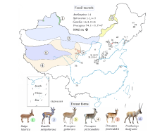 Fig. 1
Location of Shanshenmiaozui (SSMZ) site, with distributional map of living antelopes and related fossil forms of the Pleistocene Epoch in China Data for the extant taxa are from Jiang (2004), Smith and Xie (2008). Fossil localities: 1. Xiashagou of Nihewan; 2. Yushe; 3. Dongdoubi of Yuxian; 4. Jinyuan Cave; 5. Zhoukoudian Loc.1;6. Xujiayao; 7. Mashandong; 8. Salawusu; 9. Xifeng of Qingyang; 10. Loufangzi; 11. Jinniushan;12. Miaohoushan; 13. Haimao of Dalian; 14. Gulongshan; 15. Xiaogushan; 16. Chicheng; 17. Dingcun;18. Xihoudu; 19. Tianshuigou of Dali; 20. Wenxi; 21. Pinglu; 22. Danangou of Yuxian; 23. Bajiazui;24. Gonghe; 25. Yangguo; 26. Tuozidong; 27. Tunliu; 28. Banpo; 29. Linyi; 30. Yuanmou; 31. Heshui;32. Xingtai; 33. Xiaochangcun; 34. Wuwang of Linyi; 35. Gengjiagou; 36. Luhuo; 37. Zhoukoudian Loc.15;38. Upper Cave; 39. Guxiangtun; 40. Zhoujiayoufang; 41. Dali Man site; 42. Aba; 43. Chifeng;44. Rouyuan; 45. Yanjiagang; 46. Yuhongcun of Dali; 47. Lingjing
Fig. 1
Location of Shanshenmiaozui (SSMZ) site, with distributional map of living antelopes and related fossil forms of the Pleistocene Epoch in China Data for the extant taxa are from Jiang (2004), Smith and Xie (2008). Fossil localities: 1. Xiashagou of Nihewan; 2. Yushe; 3. Dongdoubi of Yuxian; 4. Jinyuan Cave; 5. Zhoukoudian Loc.1;6. Xujiayao; 7. Mashandong; 8. Salawusu; 9. Xifeng of Qingyang; 10. Loufangzi; 11. Jinniushan;12. Miaohoushan; 13. Haimao of Dalian; 14. Gulongshan; 15. Xiaogushan; 16. Chicheng; 17. Dingcun;18. Xihoudu; 19. Tianshuigou of Dali; 20. Wenxi; 21. Pinglu; 22. Danangou of Yuxian; 23. Bajiazui;24. Gonghe; 25. Yangguo; 26. Tuozidong; 27. Tunliu; 28. Banpo; 29. Linyi; 30. Yuanmou; 31. Heshui;32. Xingtai; 33. Xiaochangcun; 34. Wuwang of Linyi; 35. Gengjiagou; 36. Luhuo; 37. Zhoukoudian Loc.15;38. Upper Cave; 39. Guxiangtun; 40. Zhoujiayoufang; 41. Dali Man site; 42. Aba; 43. Chifeng;44. Rouyuan; 45. Yanjiagang; 46. Yuhongcun of Dali; 47. Lingjing
-
 Table 1
Studied fossil specimens of small to medium-sized bovids newly unearthed from SSMZ
Table 1
Studied fossil specimens of small to medium-sized bovids newly unearthed from SSMZ
-
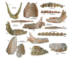 Fig. 2
Partial skulls, jaw bones and teeth of Spirocerus wongi (A-D) and Ovis shantungensis (E) from SSMZ A-D. Spirocerus wongi: A. partial skull with horn-cores (IVPP V 28650), B1-B3. juvenile maxilla with DP2-M1 (V 28651), C1-C4. left mandible with dp2-m1 (V 28652.1), D1-D4. partial right mandible with p4-m3 (V 28653); E1-E2. Ovis shantungensis, maxilla with left DP4-M1 and right DP2-4 and M1 (V 28693) A. anterior view; B1, E1. palatal views; B2, C1, D1, E2. buccal views; C2, D2. lingual views;B3, C3-4, D3-4. occlusal views. The unmarked scale bars equal 20 mm
Fig. 2
Partial skulls, jaw bones and teeth of Spirocerus wongi (A-D) and Ovis shantungensis (E) from SSMZ A-D. Spirocerus wongi: A. partial skull with horn-cores (IVPP V 28650), B1-B3. juvenile maxilla with DP2-M1 (V 28651), C1-C4. left mandible with dp2-m1 (V 28652.1), D1-D4. partial right mandible with p4-m3 (V 28653); E1-E2. Ovis shantungensis, maxilla with left DP4-M1 and right DP2-4 and M1 (V 28693) A. anterior view; B1, E1. palatal views; B2, C1, D1, E2. buccal views; C2, D2. lingual views;B3, C3-4, D3-4. occlusal views. The unmarked scale bars equal 20 mm
-
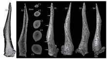 Fig. 3
CT scan images and 3-D reconstructions of the horn-core of Spirocerus wongi from SSMZ (IVPP V 28650) A1-A2. CT scan images showing the general canal system (A1) and a longitudinal slice (A2);C1-C5. CT scan slices showing the changes of cross sections and the canal system at different levels;B1-B4. CT image reconstruction of the right horn-core in anterior (B1), lateral (B2), posterior (B3) and medial (B4) views
Fig. 3
CT scan images and 3-D reconstructions of the horn-core of Spirocerus wongi from SSMZ (IVPP V 28650) A1-A2. CT scan images showing the general canal system (A1) and a longitudinal slice (A2);C1-C5. CT scan slices showing the changes of cross sections and the canal system at different levels;B1-B4. CT image reconstruction of the right horn-core in anterior (B1), lateral (B2), posterior (B3) and medial (B4) views
-
Table 2
Measurements of the horn-cores of Spirocerus wongi, compared with related species (mm)
-
Table 3
Measurements of the teeth of Spirocerus wongi, compared with related species (mm)
-
Table 4
Measurements of metacarpals of bovids from Nihewan Basin, compared with related taxa (mm)
-
 Fig. 5
Incomplete skulls and horn-cores of Gazella sinensis from SSMZ A. partial skull of a juvenile with horn-cores (IVPP V 28658); B1-B3. partial skull with horn-cores (V 28657); C1-C2. left and right horn-cores (V 28659.1, V 28659.2); D1-D2. partial skull with left horn-core (V 28667);E. partial skull with right horn-core (V 28661). A, B1, C1-2, D1. anterior views; B3. posterior view;B2. dorsal view; E. medial view; D2. lateral view. Scale bars equal 50 mm
Fig. 5
Incomplete skulls and horn-cores of Gazella sinensis from SSMZ A. partial skull of a juvenile with horn-cores (IVPP V 28658); B1-B3. partial skull with horn-cores (V 28657); C1-C2. left and right horn-cores (V 28659.1, V 28659.2); D1-D2. partial skull with left horn-core (V 28667);E. partial skull with right horn-core (V 28661). A, B1, C1-2, D1. anterior views; B3. posterior view;B2. dorsal view; E. medial view; D2. lateral view. Scale bars equal 50 mm
-
Table 5
Measurements of partial cranial bones and horn-cores of Gazella sinensis (mm)
-
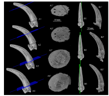 Fig. 6
CT scan images of the horn-core of Gazella sinensis (IVPP V 28661) from SSMZ A1-A4. CT image reconstruction showing positions of the cross CT scan slices (A1’-A4’);B1-B3. CT image reconstruction showing positions of the longitudinal CT scan slices (B1’-B3’)
Fig. 6
CT scan images of the horn-core of Gazella sinensis (IVPP V 28661) from SSMZ A1-A4. CT image reconstruction showing positions of the cross CT scan slices (A1’-A4’);B1-B3. CT image reconstruction showing positions of the longitudinal CT scan slices (B1’-B3’)
-
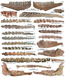 Fig. 7
Jaw bones and teeth of Gazella sinensis from SSMZ (A-J), compared with related taxa (K-P) A. maxilla with P3-M1 (IVPP V 28673); B. maxilla with M1-3 (V 28674);C1-C4. left mandible with p2-m3 (V 28680); D1-D3. left mandible with p2-m3 (V 28681.2);E1-E2. left mandible with dp2-4 and m1-2 (V 28685.1), E3. detail of dp2-4; F. right p4-m3 (V 28677);G. left p3-m3 (V 28682); H. right p2-m3 (V 28675); I. left p4-m3 (V 28686); J. left p4-m3 (V 28678);K. Procapra przewalskii, right p2-m3 (horizontally flipped) (NWIPB-0001172♀);L. Procapra gutturosa, left p2-m3 (NWIPB 620032); M. Gazella subgutturosa, left p2-m3 (NWIPB 609001);N. Pseudois nahaur, left p2-m3 (NWIPB-KX1); O. Capra ibex, left p2-m3 (IOZ-2);P. Ovis ammon, left p2-m3 (OV 1346-2). A, B, C1, C4, D1, E3, F-P. occlusal views;C2, D2, E1. buccal views; C3, D3, E2. lingual views. The arrows indicate the variations of p4 The unmarked scale bars equal 20 mm
Fig. 7
Jaw bones and teeth of Gazella sinensis from SSMZ (A-J), compared with related taxa (K-P) A. maxilla with P3-M1 (IVPP V 28673); B. maxilla with M1-3 (V 28674);C1-C4. left mandible with p2-m3 (V 28680); D1-D3. left mandible with p2-m3 (V 28681.2);E1-E2. left mandible with dp2-4 and m1-2 (V 28685.1), E3. detail of dp2-4; F. right p4-m3 (V 28677);G. left p3-m3 (V 28682); H. right p2-m3 (V 28675); I. left p4-m3 (V 28686); J. left p4-m3 (V 28678);K. Procapra przewalskii, right p2-m3 (horizontally flipped) (NWIPB-0001172♀);L. Procapra gutturosa, left p2-m3 (NWIPB 620032); M. Gazella subgutturosa, left p2-m3 (NWIPB 609001);N. Pseudois nahaur, left p2-m3 (NWIPB-KX1); O. Capra ibex, left p2-m3 (IOZ-2);P. Ovis ammon, left p2-m3 (OV 1346-2). A, B, C1, C4, D1, E3, F-P. occlusal views;C2, D2, E1. buccal views; C3, D3, E2. lingual views. The arrows indicate the variations of p4 The unmarked scale bars equal 20 mm
-
Table 6
Measurements of teeth of Gazella sinensis from SSMZ, compared with those from Xiashagou (mm)
-
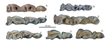 Fig. 8
Comparison of premolar series among some Quaternary gazelle species of China A. Gazella sinensis, left p2-4, IVPP V 28681.2, SSMZ; B. Procapra gutturosa, left p2-4, NWIPB 0006065, extant; C. P. przewalskii, right p2-4 (horizontally flipped), NWIPB 0001172, extant;D. P. picticaudata, left p2-4, NWIPB 0001179, extant; E. Gazella subgutturosa, left p2-4, IVPP-c-05, extant;F. Saiga tatarica, right p3-4 (horizontally flipped), NWIPB S-80503, extant;G. Pantholops hodgsonii, left p3-4, NWIPB 77001, extant. All are in occlusal views
Fig. 8
Comparison of premolar series among some Quaternary gazelle species of China A. Gazella sinensis, left p2-4, IVPP V 28681.2, SSMZ; B. Procapra gutturosa, left p2-4, NWIPB 0006065, extant; C. P. przewalskii, right p2-4 (horizontally flipped), NWIPB 0001172, extant;D. P. picticaudata, left p2-4, NWIPB 0001179, extant; E. Gazella subgutturosa, left p2-4, IVPP-c-05, extant;F. Saiga tatarica, right p3-4 (horizontally flipped), NWIPB S-80503, extant;G. Pantholops hodgsonii, left p3-4, NWIPB 77001, extant. All are in occlusal views
-
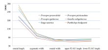 Fig. 9
Toothrow length and cranial size of extant gazelles in China Raw data was employed from Jiang, 2004
Fig. 9
Toothrow length and cranial size of extant gazelles in China Raw data was employed from Jiang, 2004
-
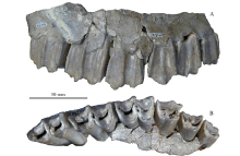 Fig. 10
Maxilla of Megalovis piveteaui (MNHN-NIH 150) from Xiashagou of Nihewan A. buccal view; B. occlusal view
Fig. 10
Maxilla of Megalovis piveteaui (MNHN-NIH 150) from Xiashagou of Nihewan A. buccal view; B. occlusal view
-
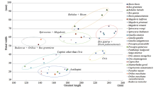 Fig. 11
Length vs distal width of metacarpal bones of diverse bovids The data un-included in Table 4 are from Colbert and Hooijer, 1953; Scott, 1985
Fig. 11
Length vs distal width of metacarpal bones of diverse bovids The data un-included in Table 4 are from Colbert and Hooijer, 1953; Scott, 1985
|
