New fossils of small and medium-sized bovids from the Early Site of Shanshenmiaozui in Nihewan Basin, North China
TONG Hao-Wen, ZHANG Bei, CHEN Xi, WANG Xiao-Min
Vertebrata Palasiatica
2022, 60 ( 2):
134-168.
DOI: 10.19615/j.cnki.2096-9899.220413
Shanshenmiaozui site in Nihewan Basin in North China is a recently discovered Early Pleistocene site which yields rich and diverse mammalian fossils. In the fauna, the small and medium-sized bovid fossils are well represented and can be referred to the following taxa: Spirocerus wongi, Gazella sinensis, Ovis shantungensis and Megalovis piveteaui respectively, among which G. sinensis is the dominate species. S. wongi and G. sinensis are mainly represented by horn-cores and partial skull bones as well as mandibles; in addition, metacarpal and/or metatarsal bones were also recognized for all of the four species. The horn-cores are easy to be identified to the species level, while the dentitions and the postcranial bones underwent a series of examinations and comparisons before getting properly determined and referred to the most approximate taxa. Among the postcranial bones, the metapodials, especially to the metacarpal bones special attentions were paid, which are crucial not only for taxonomic identification, but also for phylogenetic and paleoecological reconstructions; the previously misidentified metapodial specimens in Nihewan fauna were reconsidered in this paper. In the SSMZ fauna, the bovid guild is dominated by Gazella and Bison, which indicates steppe was the most important biome in Nihewan Basin during Early Pleistocene.
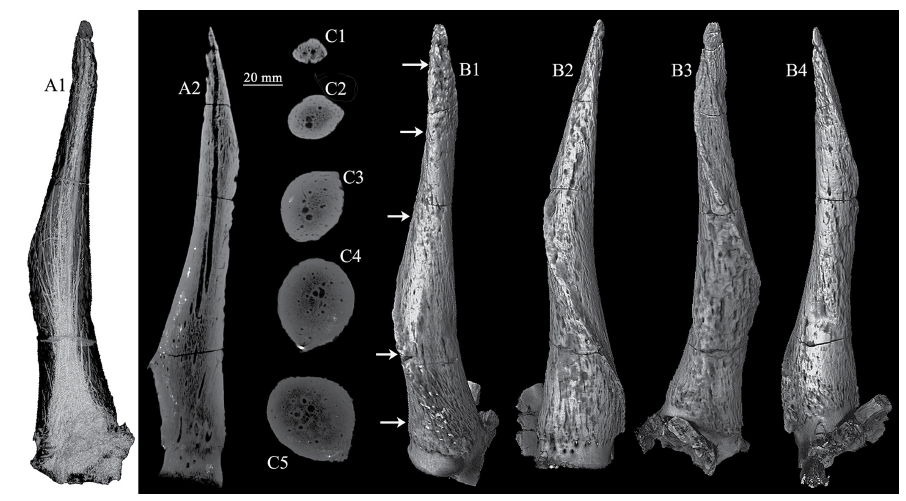
Fig. 3
CT scan images and 3-D reconstructions of the horn-core of Spirocerus wongi from SSMZ (IVPP V 28650)
A1-A2. CT scan images showing the general canal system (A1) and a longitudinal slice (A2);C1-C5. CT scan slices showing the changes of cross sections and the canal system at different levels;B1-B4. CT image reconstruction of the right horn-core in anterior (B1), lateral (B2), posterior (B3) and medial (B4) views
Extracts from the Article
Skull bones: One partial adult skull (Fig. 2A) and one partial juvenile skull (Fig. 2B1-3) can be referred to the the species S. wongi. The adult one has very limited frontal bone, but with almost complete horn-cores of both sides. In front view, the frontal bone shows two moderately developed supraorbital pits which accommodate the two prominent supraorbital foramina which are connecting directly to the anterosuperior corner of the orbit and each has a very large internal opening, even larger than in an ox. The eye socket is very large and deep, and has a trench-like roof with flat superior wall rather than domed as in other taxa. The horn is very close to the orbit, which is only 30 mm apart; the postorbital constriction is also obvious. The cranial width at the horn bases is 149.0 mm, and the two horn-cores are 65.6 mm apart at the base. The frontal sinus (pneumatization) is also quite developed, but the diverticulum doesn’t extend to the cornual process (Fig. 3A2).
Horn-core: The horn-core is a typical form of Spirocerus wongi, with rugose surface and only one spiral carena (or carène or keel) (Fig. 3B1-4), rather than two as in S. peii and S. kiakhtensis. The carena derives from the basal anterior part and heteronymously spirals upwardly (the left horn spirals anticlockwise but the right one spirals clockwise) and ends near the tip after finishing one complete revolution. The pedicle is quite short. The interior of the horn-core is nearly solid but with a complicated minor canal system (Fig. 3A1-2, C1-5), which resembles those of the true gazelles. The cross-section of the horn-core is elliptical (Fig. 3C1-5). Around the basal part of the horn-core, there are many nutrient foramina, among which the ones above the orbit are the largest (Fig. 3B2). The dimensions of the horn-cores are shown in Table 2.
Spirocerus has developed frontal sinuses, but they do not extend to the horn-core at all (Fig. 3A2). According to Farke’s paper of 2010, the frontal sinus doesn’t always have phylogenetic significance. But the present authors think it’s more important if the frontal sinus extends to the horn-core, i.e. if there is a cornual diverticulum of the frontal sinus or if the horn-core is hollowed.
Other Images/Table from this Article
-
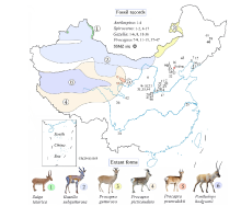 Fig. 1
Location of Shanshenmiaozui (SSMZ) site, with distributional map of living antelopes and related fossil forms of the Pleistocene Epoch in China Data for the extant taxa are from Jiang (2004), Smith and Xie (2008). Fossil localities: 1. Xiashagou of Nihewan; 2. Yushe; 3. Dongdoubi of Yuxian; 4. Jinyuan Cave; 5. Zhoukoudian Loc.1;6. Xujiayao; 7. Mashandong; 8. Salawusu; 9. Xifeng of Qingyang; 10. Loufangzi; 11. Jinniushan;12. Miaohoushan; 13. Haimao of Dalian; 14. Gulongshan; 15. Xiaogushan; 16. Chicheng; 17. Dingcun;18. Xihoudu; 19. Tianshuigou of Dali; 20. Wenxi; 21. Pinglu; 22. Danangou of Yuxian; 23. Bajiazui;24. Gonghe; 25. Yangguo; 26. Tuozidong; 27. Tunliu; 28. Banpo; 29. Linyi; 30. Yuanmou; 31. Heshui;32. Xingtai; 33. Xiaochangcun; 34. Wuwang of Linyi; 35. Gengjiagou; 36. Luhuo; 37. Zhoukoudian Loc.15;38. Upper Cave; 39. Guxiangtun; 40. Zhoujiayoufang; 41. Dali Man site; 42. Aba; 43. Chifeng;44. Rouyuan; 45. Yanjiagang; 46. Yuhongcun of Dali; 47. Lingjing
Fig. 1
Location of Shanshenmiaozui (SSMZ) site, with distributional map of living antelopes and related fossil forms of the Pleistocene Epoch in China Data for the extant taxa are from Jiang (2004), Smith and Xie (2008). Fossil localities: 1. Xiashagou of Nihewan; 2. Yushe; 3. Dongdoubi of Yuxian; 4. Jinyuan Cave; 5. Zhoukoudian Loc.1;6. Xujiayao; 7. Mashandong; 8. Salawusu; 9. Xifeng of Qingyang; 10. Loufangzi; 11. Jinniushan;12. Miaohoushan; 13. Haimao of Dalian; 14. Gulongshan; 15. Xiaogushan; 16. Chicheng; 17. Dingcun;18. Xihoudu; 19. Tianshuigou of Dali; 20. Wenxi; 21. Pinglu; 22. Danangou of Yuxian; 23. Bajiazui;24. Gonghe; 25. Yangguo; 26. Tuozidong; 27. Tunliu; 28. Banpo; 29. Linyi; 30. Yuanmou; 31. Heshui;32. Xingtai; 33. Xiaochangcun; 34. Wuwang of Linyi; 35. Gengjiagou; 36. Luhuo; 37. Zhoukoudian Loc.15;38. Upper Cave; 39. Guxiangtun; 40. Zhoujiayoufang; 41. Dali Man site; 42. Aba; 43. Chifeng;44. Rouyuan; 45. Yanjiagang; 46. Yuhongcun of Dali; 47. Lingjing
-
 Table 1
Studied fossil specimens of small to medium-sized bovids newly unearthed from SSMZ
Table 1
Studied fossil specimens of small to medium-sized bovids newly unearthed from SSMZ
-
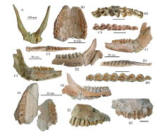 Fig. 2
Partial skulls, jaw bones and teeth of Spirocerus wongi (A-D) and Ovis shantungensis (E) from SSMZ A-D. Spirocerus wongi: A. partial skull with horn-cores (IVPP V 28650), B1-B3. juvenile maxilla with DP2-M1 (V 28651), C1-C4. left mandible with dp2-m1 (V 28652.1), D1-D4. partial right mandible with p4-m3 (V 28653); E1-E2. Ovis shantungensis, maxilla with left DP4-M1 and right DP2-4 and M1 (V 28693) A. anterior view; B1, E1. palatal views; B2, C1, D1, E2. buccal views; C2, D2. lingual views;B3, C3-4, D3-4. occlusal views. The unmarked scale bars equal 20 mm
Fig. 2
Partial skulls, jaw bones and teeth of Spirocerus wongi (A-D) and Ovis shantungensis (E) from SSMZ A-D. Spirocerus wongi: A. partial skull with horn-cores (IVPP V 28650), B1-B3. juvenile maxilla with DP2-M1 (V 28651), C1-C4. left mandible with dp2-m1 (V 28652.1), D1-D4. partial right mandible with p4-m3 (V 28653); E1-E2. Ovis shantungensis, maxilla with left DP4-M1 and right DP2-4 and M1 (V 28693) A. anterior view; B1, E1. palatal views; B2, C1, D1, E2. buccal views; C2, D2. lingual views;B3, C3-4, D3-4. occlusal views. The unmarked scale bars equal 20 mm
-
Table 2
Measurements of the horn-cores of Spirocerus wongi, compared with related species (mm)
-
Table 3
Measurements of the teeth of Spirocerus wongi, compared with related species (mm)
-
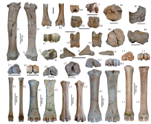 Fig. 4
Postcranial bones of small and medium-sized bovids from SSMZ A-C. Spirocerus wongi: A1-A3. left Mc III+IV (IVPP V 28655),B1-B4. right naviculo-cuboid+lat-mid+medial cuneiforms (V 28656.2-3),C1-C3. right Mt III+IV (V 28656.4); D-I. Gazella sinensis: D1-D3. partial left humerus (V 28687),E1-E4. left Mc III+IV (V 28688), F. distal epiphysis of left tibia (V 28691),G1-G2. left astragalus (V 28692), H1-H2. left naviculo-cuboid (V 28690), I1-I3. 3rd phalanx (V 28689);J-K. Megalovis piveteaui: J1-J4. left radius (V 28654), K1-K4. right Mc III+IV (V 28695);L1-L4. Ovis shantungensis: left Mc III+IV (V 28694). A1, C1, D1, E1, G1, J1, K1, L1. anterior views;A2, B4, C2, D2, E2, G2, J2, K2, L2. posterior views; A3, B1, C3, E3, H1, J3, K3, L3. proximal views;B2, D3, E4, F, H2, J4, K4, L4. distal views; B3. medial view; I1. lateral view; I2. interdigital view;I3. volar view. The arrows indicate the lateral projection of the radius. The unmarked scale bars equal 20 mm
Fig. 4
Postcranial bones of small and medium-sized bovids from SSMZ A-C. Spirocerus wongi: A1-A3. left Mc III+IV (IVPP V 28655),B1-B4. right naviculo-cuboid+lat-mid+medial cuneiforms (V 28656.2-3),C1-C3. right Mt III+IV (V 28656.4); D-I. Gazella sinensis: D1-D3. partial left humerus (V 28687),E1-E4. left Mc III+IV (V 28688), F. distal epiphysis of left tibia (V 28691),G1-G2. left astragalus (V 28692), H1-H2. left naviculo-cuboid (V 28690), I1-I3. 3rd phalanx (V 28689);J-K. Megalovis piveteaui: J1-J4. left radius (V 28654), K1-K4. right Mc III+IV (V 28695);L1-L4. Ovis shantungensis: left Mc III+IV (V 28694). A1, C1, D1, E1, G1, J1, K1, L1. anterior views;A2, B4, C2, D2, E2, G2, J2, K2, L2. posterior views; A3, B1, C3, E3, H1, J3, K3, L3. proximal views;B2, D3, E4, F, H2, J4, K4, L4. distal views; B3. medial view; I1. lateral view; I2. interdigital view;I3. volar view. The arrows indicate the lateral projection of the radius. The unmarked scale bars equal 20 mm
-
Table 4
Measurements of metacarpals of bovids from Nihewan Basin, compared with related taxa (mm)
-
 Fig. 5
Incomplete skulls and horn-cores of Gazella sinensis from SSMZ A. partial skull of a juvenile with horn-cores (IVPP V 28658); B1-B3. partial skull with horn-cores (V 28657); C1-C2. left and right horn-cores (V 28659.1, V 28659.2); D1-D2. partial skull with left horn-core (V 28667);E. partial skull with right horn-core (V 28661). A, B1, C1-2, D1. anterior views; B3. posterior view;B2. dorsal view; E. medial view; D2. lateral view. Scale bars equal 50 mm
Fig. 5
Incomplete skulls and horn-cores of Gazella sinensis from SSMZ A. partial skull of a juvenile with horn-cores (IVPP V 28658); B1-B3. partial skull with horn-cores (V 28657); C1-C2. left and right horn-cores (V 28659.1, V 28659.2); D1-D2. partial skull with left horn-core (V 28667);E. partial skull with right horn-core (V 28661). A, B1, C1-2, D1. anterior views; B3. posterior view;B2. dorsal view; E. medial view; D2. lateral view. Scale bars equal 50 mm
-
Table 5
Measurements of partial cranial bones and horn-cores of Gazella sinensis (mm)
-
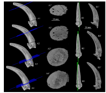 Fig. 6
CT scan images of the horn-core of Gazella sinensis (IVPP V 28661) from SSMZ A1-A4. CT image reconstruction showing positions of the cross CT scan slices (A1’-A4’);B1-B3. CT image reconstruction showing positions of the longitudinal CT scan slices (B1’-B3’)
Fig. 6
CT scan images of the horn-core of Gazella sinensis (IVPP V 28661) from SSMZ A1-A4. CT image reconstruction showing positions of the cross CT scan slices (A1’-A4’);B1-B3. CT image reconstruction showing positions of the longitudinal CT scan slices (B1’-B3’)
-
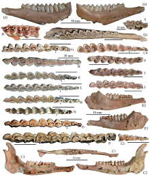 Fig. 7
Jaw bones and teeth of Gazella sinensis from SSMZ (A-J), compared with related taxa (K-P) A. maxilla with P3-M1 (IVPP V 28673); B. maxilla with M1-3 (V 28674);C1-C4. left mandible with p2-m3 (V 28680); D1-D3. left mandible with p2-m3 (V 28681.2);E1-E2. left mandible with dp2-4 and m1-2 (V 28685.1), E3. detail of dp2-4; F. right p4-m3 (V 28677);G. left p3-m3 (V 28682); H. right p2-m3 (V 28675); I. left p4-m3 (V 28686); J. left p4-m3 (V 28678);K. Procapra przewalskii, right p2-m3 (horizontally flipped) (NWIPB-0001172♀);L. Procapra gutturosa, left p2-m3 (NWIPB 620032); M. Gazella subgutturosa, left p2-m3 (NWIPB 609001);N. Pseudois nahaur, left p2-m3 (NWIPB-KX1); O. Capra ibex, left p2-m3 (IOZ-2);P. Ovis ammon, left p2-m3 (OV 1346-2). A, B, C1, C4, D1, E3, F-P. occlusal views;C2, D2, E1. buccal views; C3, D3, E2. lingual views. The arrows indicate the variations of p4 The unmarked scale bars equal 20 mm
Fig. 7
Jaw bones and teeth of Gazella sinensis from SSMZ (A-J), compared with related taxa (K-P) A. maxilla with P3-M1 (IVPP V 28673); B. maxilla with M1-3 (V 28674);C1-C4. left mandible with p2-m3 (V 28680); D1-D3. left mandible with p2-m3 (V 28681.2);E1-E2. left mandible with dp2-4 and m1-2 (V 28685.1), E3. detail of dp2-4; F. right p4-m3 (V 28677);G. left p3-m3 (V 28682); H. right p2-m3 (V 28675); I. left p4-m3 (V 28686); J. left p4-m3 (V 28678);K. Procapra przewalskii, right p2-m3 (horizontally flipped) (NWIPB-0001172♀);L. Procapra gutturosa, left p2-m3 (NWIPB 620032); M. Gazella subgutturosa, left p2-m3 (NWIPB 609001);N. Pseudois nahaur, left p2-m3 (NWIPB-KX1); O. Capra ibex, left p2-m3 (IOZ-2);P. Ovis ammon, left p2-m3 (OV 1346-2). A, B, C1, C4, D1, E3, F-P. occlusal views;C2, D2, E1. buccal views; C3, D3, E2. lingual views. The arrows indicate the variations of p4 The unmarked scale bars equal 20 mm
-
Table 6
Measurements of teeth of Gazella sinensis from SSMZ, compared with those from Xiashagou (mm)
-
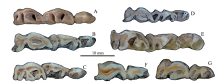 Fig. 8
Comparison of premolar series among some Quaternary gazelle species of China A. Gazella sinensis, left p2-4, IVPP V 28681.2, SSMZ; B. Procapra gutturosa, left p2-4, NWIPB 0006065, extant; C. P. przewalskii, right p2-4 (horizontally flipped), NWIPB 0001172, extant;D. P. picticaudata, left p2-4, NWIPB 0001179, extant; E. Gazella subgutturosa, left p2-4, IVPP-c-05, extant;F. Saiga tatarica, right p3-4 (horizontally flipped), NWIPB S-80503, extant;G. Pantholops hodgsonii, left p3-4, NWIPB 77001, extant. All are in occlusal views
Fig. 8
Comparison of premolar series among some Quaternary gazelle species of China A. Gazella sinensis, left p2-4, IVPP V 28681.2, SSMZ; B. Procapra gutturosa, left p2-4, NWIPB 0006065, extant; C. P. przewalskii, right p2-4 (horizontally flipped), NWIPB 0001172, extant;D. P. picticaudata, left p2-4, NWIPB 0001179, extant; E. Gazella subgutturosa, left p2-4, IVPP-c-05, extant;F. Saiga tatarica, right p3-4 (horizontally flipped), NWIPB S-80503, extant;G. Pantholops hodgsonii, left p3-4, NWIPB 77001, extant. All are in occlusal views
-
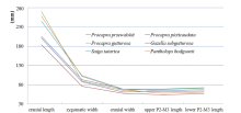 Fig. 9
Toothrow length and cranial size of extant gazelles in China Raw data was employed from Jiang, 2004
Fig. 9
Toothrow length and cranial size of extant gazelles in China Raw data was employed from Jiang, 2004
-
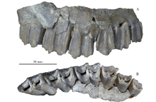 Fig. 10
Maxilla of Megalovis piveteaui (MNHN-NIH 150) from Xiashagou of Nihewan A. buccal view; B. occlusal view
Fig. 10
Maxilla of Megalovis piveteaui (MNHN-NIH 150) from Xiashagou of Nihewan A. buccal view; B. occlusal view
-
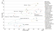 Fig. 11
Length vs distal width of metacarpal bones of diverse bovids The data un-included in Table 4 are from Colbert and Hooijer, 1953; Scott, 1985
Fig. 11
Length vs distal width of metacarpal bones of diverse bovids The data un-included in Table 4 are from Colbert and Hooijer, 1953; Scott, 1985
|
