Please wait a minute...
Welcome to Visited Vertebrata Palasiatica, Today is Share:
IMAGE/TABLE DETAILS
The first description of Rhinocerotidae (Perissodactyla, Mammalia) from Xinyaozi Ravine in Shanxi, North China
DONG Wei, BAI Wei-Peng, ZHANG Li-Min
Vertebrata Palasiatica    2021, 59 (4): 273-294.   DOI: 10.19615/j.cnki.2096-9899.210715
Abstract   (585 HTML79 PDF(pc) (3880KB)(499)  

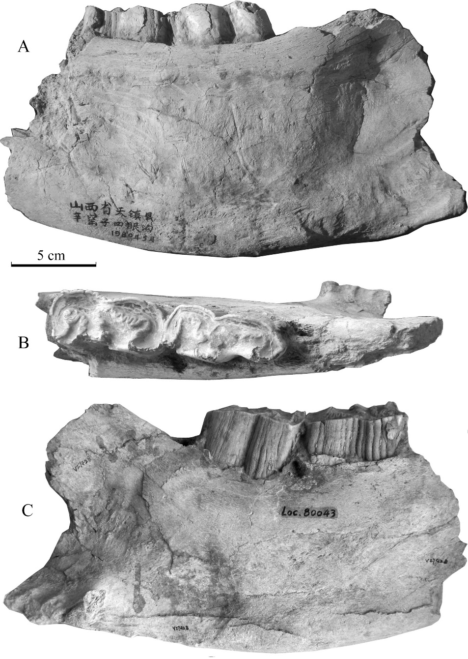
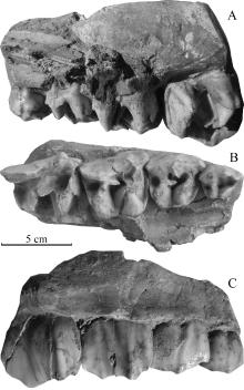
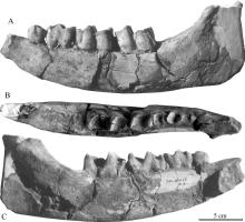
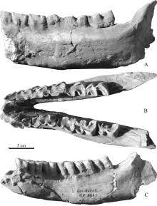
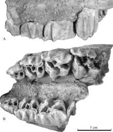
Abundant mammalian fossils were uncovered during the field exploration for Nihewan beds at the beginning of the 1980s along Xinyaozi Ravine at Nangaoya Township of Tianzhen County, Shanxi Province in North China and the studied taxa indicate an age of the early Early Pleistocene. Recent studies on the rhino material not yet described show that there are at least two species of rhinocerotids: Elasmotherium peii and Coelodonta nihowanensis . There might be a third taxon provisionally named as Stephanorhinus cf. S. kirchbergensis due to incompleteness of the specimens. Since its morphometric characters are between S. kirchbergensis and C. nihowanensis , it might be a variety of one of the two species although it is more similar to the former than the latter. In the same way, The rhino specimens from Xiashagou named as Rhinoceros sinensis (?) by Teilhard de Chardin and Piveteau (1930) might be a variety of S. kirchbergensis or C. nihowanensis . The rhinocerotids uncovered so far from the Early Pleistocene deposits in the generalized Nihewan Basin including two certain species and two uncertain ones. The localities yielding E. peii include Xiashagou, Shanshenmiaozhui, Daheigou and Xinyaozi; those yielding C. nihowanensis include Xiashagou, Danangou, Donggutuo, Shanshenmiaozhui and Xinyaozi. R. sinensis (?) appeared only at Xiashagou and Stephanorhinus cf. S. kirchbergensis only at Xinyaozi.


Fig. 1 A mandibular fragment (IVPP V 27428) of Elasmotherium peii from Xinyaozi Ravine in lingual (A), occlusal (B) and buccal (C) views
Extracts from the Article
(Fig. 1; Table 1)
Description The specimen V 27428 (Fig. 1) is a right adult mandibular fragment with m2-3. The preserved fragment indicates that the mandibular body is massive, deep (high) but not very thick. The part in front of the anterior fracture just below the anterior border of m2 of the mandibular body is broken off. The lingual side of the body is slightly convex, and its buccal side is somewhat concave. The ventral margin on the buccal side of the body forms a protrusion similar to a horizontal torus. The depth or height from the alveolar margin to ventral one of the mandibular body measured at m2 is 141.18 mm on buccal side and 153.38 mm on lingual one. And those at m3 are 154.46 and 159.92 mm respectively. The mandibular ramus is partially preserved. The masseteric tuberosity (tuberositas masseterica) is well developed and protrudes laterally (Fig. 1B-C), indicating the powerful muscle of mastication. The mastication fossa (fossa masseterica) is large and somewhat profound, and it extends to the posterior lobe of m3. The pterygoid fossa (fossa pterygoidea) is also deep but less large and just extends to the posterior border of m3. The rest parts of the mandibular ramus, such as coronoid process, condyle process, angular process and mandibular angle are all missing. The specimen is heavily mineralized and resistant to the X ray of CT scanner and the slices obtained are not very clear.
Other Images/Table from this Article